Coronary angiogram
An X-ray of the heart arteries, done under local anaesthetic.
At a glance
- How long
- Usually about 30 minutes, and you go home the same day
- Anaesthetic
- Local, at the wrist or groin, and you stay awake
- Getting home
- Arrange for someone to take you home
- Recovery
- Most normal activities the next day
What the test involves
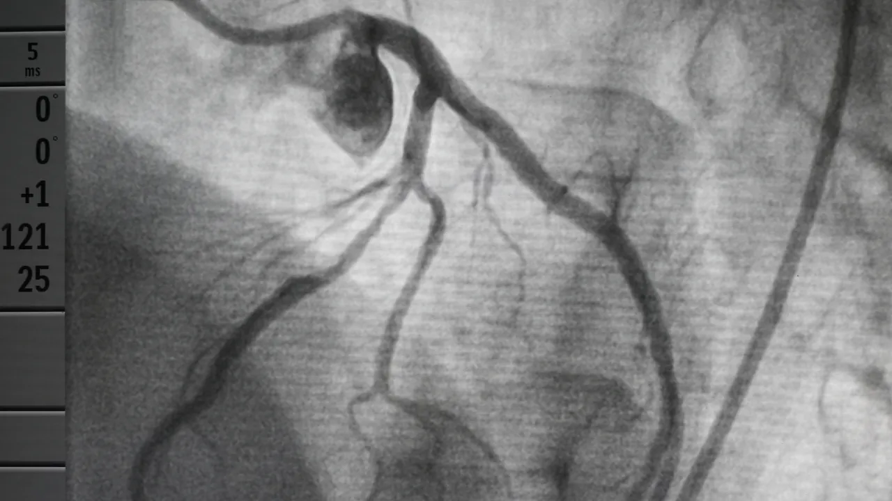
This is a test where the heart arteries can be directly visualised by an X-ray. Under local anaesthetic a tube is passed via the artery in the wrist or leg into the heart, and dye injected into the heart arteries. This is still the best way of establishing whether there is any narrowing of the heart arteries.
Tests are charged by the hospital that carries them out.
Why you might need a coronary angiogram
A coronary angiogram shows narrowed areas or blockages in the coronary arteries, the blood vessels that supply the heart muscle, which are the cause of angina and coronary artery disease. It is still the most reliable way to see this, particularly in the smaller arteries and branches.
It is often arranged when a CT coronary angiogram or an exercise ECG suggests narrowing, or when the answer is not yet clear. If narrowing is found, it can sometimes be treated during the same procedure, by widening the artery with a small balloon and a stent (angioplasty).
Before the test
- You will be asked about your health, any allergies and your medicines, and told if any need stopping beforehand. A blood test is usual.
- Do not eat or drink for a few hours beforehand, as instructed by the hospital.
- Tell the team if you have diabetes, so they can plan for it.
- Arrange for someone to take you home. If you have a sedative, you will not be able to drive yourself.
What happens during a coronary angiogram
The test is done in a cardiac catheter laboratory, often called the cath lab. You lie flat and stay awake, so you can talk to the team, and you can ask for a mild sedative if you feel anxious. A monitor and a clip on your finger follow your heart rate, rhythm and oxygen level.
Your wrist or the top of your leg is numbed with local anaesthetic. A thin, flexible tube is passed into the artery there and guided to your heart using X-rays. You may feel some pushing, but it should not hurt. Dye is then injected into the heart arteries and X-ray pictures are taken. As the dye goes in, a hot, flushing feeling is very common, and some people briefly feel as if they need to pass urine. Both pass within a few seconds.
After the test
The tube is removed and the artery is closed off with a tight band on the wrist, or with a seal or pressure on the groin. No stitches are needed. After the wrist approach you can usually sit up in a chair almost straight away; after the groin approach you will need to lie still for longer. Drinking plenty of water helps flush the dye out.
The doctor may be able to explain what was found before you leave, although the pictures sometimes need studying in more detail first. At home, rest for the rest of the day, and it is a good idea for someone to stay with you for 24 hours. Most people return to their normal activities the next day, but avoid heavy lifting and strenuous exercise for a few days. Some bruising and soreness at the wrist or groin for a few days is normal.
Risks of a coronary angiogram
A coronary angiogram is a very common and relatively safe test, and it is only done when the benefits outweigh the risks. Bruising, soreness and a small lump where the tube went in are common and settle over days or weeks. Less often, the cut becomes infected or the dye causes a mild itchy rash.
Serious complications are very rare. They include a heart attack or stroke, damage to the artery causing bleeding, a severe allergic reaction to the dye, and kidney problems from the dye, which are usually temporary. The test also uses a small amount of radiation. Your own risks will be discussed with you beforehand.
When to get medical help afterwards
Contact your GP or the hospital if:
- the cut starts bleeding and does not stop after pressing on it for a few minutes
- you have severe pain that painkillers do not relieve
- the skin around the cut becomes red, swollen and hot
- the arm or leg used looks or feels different from the other one, for example pale or cold
- a firm lump that is sore to touch develops near the cut
The hospital that carries out your test will send you its own instructions. If they differ from this page, follow theirs, and ask if anything is unclear.
Where this information comes from
Sources checked 14 October 2026.