Echocardiogram
A painless ultrasound scan of the heart muscle and valves.
At a glance
- How long
- Usually 30 to 40 minutes
- Does it hurt?
- No, you may feel light pressure from the probe
- Preparation
- None, eat and drink as normal
- Radiation
- None, it uses sound waves
What the test involves
An echocardiogram (or cardiac ultrasound test) is a way of directly imaging the heart muscle and heart valves. It is similar to other ultrasound tests and is completely painless. It can assess how efficiently the heart is working. It can also provide information on the heart valves as well as the sac surrounding the heart (the pericardium). This test is useful in virtually every type of cardiac symptom.
Tests are charged by the hospital that carries them out.
Why you might need an echocardiogram
An echocardiogram, or echo, is an ultrasound scan of the heart, much like the scans used in pregnancy. It shows the heart's chambers and valves, how blood flows through them and how well the heart is pumping. It is often used to diagnose or keep an eye on:
- heart failure
- heart valve problems, including a heart murmur heard through a stethoscope
- damage to the heart muscle after a heart attack
- congenital heart conditions and inherited ones such as cardiomyopathy
It is a common next step for breathlessness, or after an ECG that needs a closer look.
What happens during an echocardiogram
You remove your clothing from the waist up and wear a gown, behind a curtain or in a private room. You lie on a couch, often turned on your left side, and small sticky pads are put on your chest to follow your heart rate.
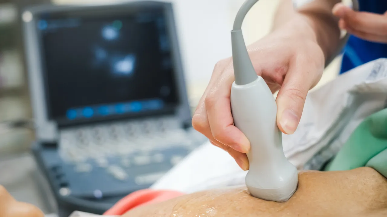
The person doing the scan, called a sonographer, spreads a cold gel on your chest and moves a small probe over it. Pictures of your heart appear on a screen. You may hear a swishing sound, which is the sound of blood flowing through the heart. Sometimes a contrast agent is injected into a vein in your arm to make the pictures clearer, and you may be asked to wait up to 30 minutes afterwards.
A standard echo has no risks or side effects, and you can go home as soon as it is finished.
Other types of echocardiogram
Dr Vasudeva has a special interest in echocardiography, both trans-thoracic (the standard scan through the chest) and trans-oesophageal. Depending on the question being asked, you may be offered:
- A transoesophageal echo (TOE). A thin probe is passed down the throat, which sits just behind the heart, to give more detailed pictures. Your throat is numbed with a spray and you can have a light sedative. It takes about 30 minutes, you may need to avoid eating for several hours beforehand, and you must not drive for 24 hours if you are sedated. A sore throat afterwards should pass within a day or two.
- A stress echo. The scan is repeated while your heart is beating faster, either through exercise or with a medicine given into a vein. It can show whether part of the heart muscle is short of blood, a sign of coronary heart disease.
What an echocardiogram cannot show
An echo does not show the coronary arteries, the blood vessels that feed the heart muscle. A CT coronary angiogram looks at those. It also cannot catch a rhythm problem that comes and goes, which needs a longer recording such as an ambulatory ECG.
The hospital that carries out your test will send you its own instructions. If they differ from this page, follow theirs, and ask if anything is unclear.
Where this information comes from
Sources checked 14 October 2026.
Symptoms this test can help investigate
- Chest painChest pain that is aching in nature, and which is brought on by exertion (walking or other exercise), is usually referred to as angina, and can be due to a narrowing of the arteries feeding the heart muscle.Read about chest pain
- PalpitationsPalpitations that feel like fluttering or pounding, and which occur at rest or with activity, are linked to common triggers but could sometimes indicate rhythm problems within the heart's electrical system.Read about palpitations
- BreathlessnessBreathlessness that develops with activity, and which feels out of proportion to your level of exertion, is often linked to heart failure, where the heart does not pump efficiently due to valve disease, rhythm problems, or previous heart damage.Read about breathlessness
- Dizziness or blackoutsDizziness that comes on suddenly, and which may be linked to brief drops in blood pressure or vaso vagal episodes, can also sometimes signal underlying rhythm problems that need assessment to ensure a pacemaker is not required.Read about dizziness or blackouts
- FatiguePersistent tiredness that is not explained by activity levels, sleep, or recent illness can sometimes be linked to heart conditions.Read about fatigue